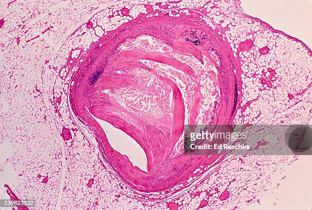
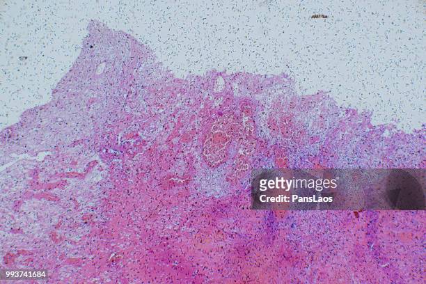
atherosclerosis of coronary artery, 5x at 35mm. reduced lumen. wall has excess calcification & fibrous connective tissue. h - microphotographie immunofluorescente photos et images de collection
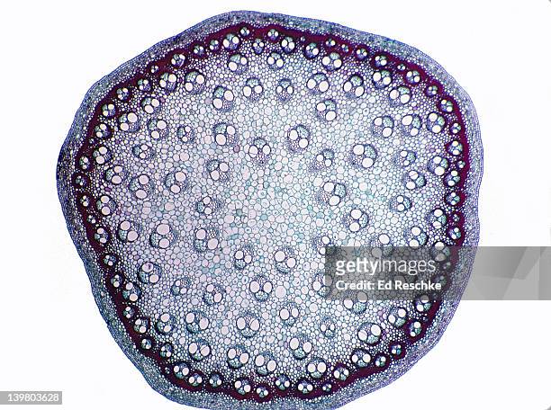
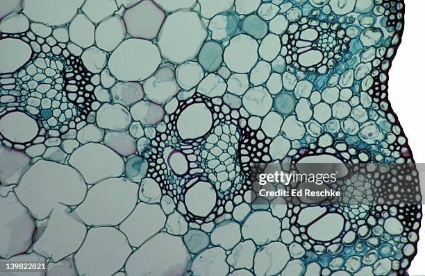
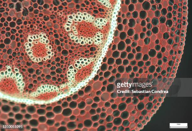
stem cross section. corn (zea), herbaceous monocot, 5x. shows: scattered vascular bundles typical of monocots, xylem, phloem, pith and epidermis. - microphotographie immunofluorescente photos et images de collection
smilax, monocot stem cross section, 8x. shows: scattered vascular bundles, xylem, phloem, cortex and epidermis. - microphotographie immunofluorescente photos et images de collection
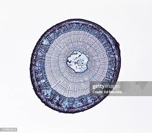
woody dicot stem. tilia (basswood), 3 year stem, 5x. shows: 3 annual rings, xylem, phloem, rays, pith, cambium, and periderm. - microphotographie immunofluorescente photos et images de collection
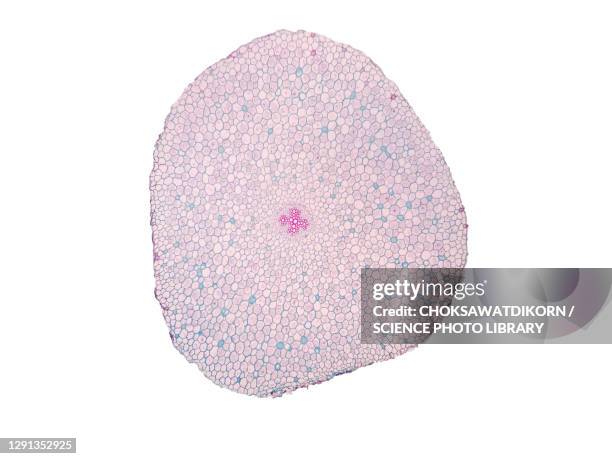
stem cross section. buttercup (ranunculus), herbaceous dicot, 8x. shows: vascular bundles arranged in a ring (typical of dicots), xylem, phloem, epidermis, cortex, and pith. - microphotographie immunofluorescente photos et images de collection
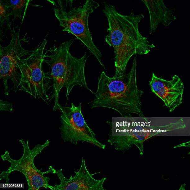
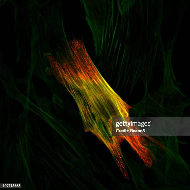
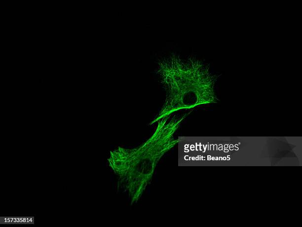
human cell under microscope. evaluated actin filaments(nanotechnology) and morphological changes following chemotherapy using confocal microscopy - microphotographie immunofluorescente photos et images de collection
simple squamous epithelium. endothelium (lining an artery) 250x. this epithelium lines the inside of blood vessels. it is extrememly flat, note flattened nuclei. also shows internal elastic membrane & smooth muscle in artery wall. - microphotographie immunofluorescente photos et images de collection
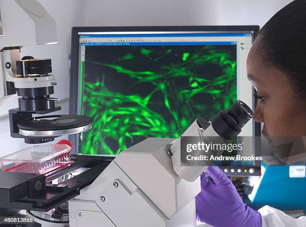
female researcher using inverted microscope to view stem cells displayed showing fluorescent labeled cells - microphotographie immunofluorescente photos et images de collection
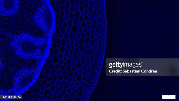
microscopic view of cells of broad bean plant. immunofluorescent photomicrograph, organs samples, histological examination, histopathology on the microscope - microphotographie immunofluorescente photos et images de collection
la microscopie en immunofluorescence - microphotographie immunofluorescente photos et images de collection
slide demonstrating breast tissue with ductal carcinoma. histopathology on the microscope. immunofluorescent photomicrograph, organs samples, histological examination, - microphotographie immunofluorescente photos et images de collection
immunofluorescent photomicrograph, organs samples, histological examination, histopathology on the microscope - microphotographie immunofluorescente photos et images de collection
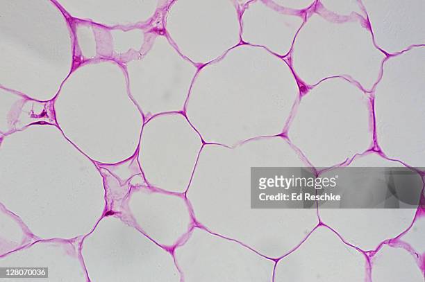
adipose tissue (magnification x 100) a specialized type of connective tissue, showing fat cells (adipocytes), nuclei (peripheral), cytoplasm, and large, clear spaces where fat was stored. - microphotographie immunofluorescente photos et images de collection
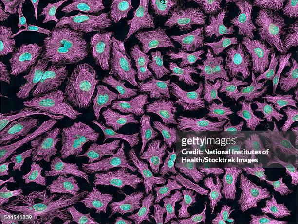
multiphoton fluorescence image of hela cells. - microphotographie immunofluorescente photos et images de collection
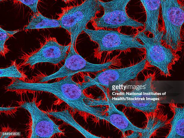
multiphoton fluorescence image of hela cells. - microphotographie immunofluorescente photos et images de collection
villus cross section--simple columnar epithelium - microphotographie immunofluorescente photos et images de collection
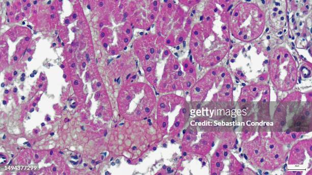
human liver tissue, light micrograph - microphotographie immunofluorescente photos et images de collection
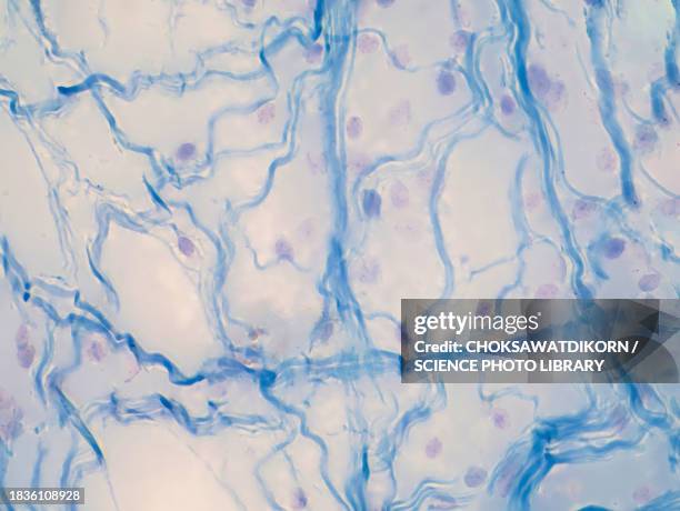
areolar connective tissue, light micrograph - microphotographie immunofluorescente photos et images de collection
microscopic photo of a professionally prepared slide demonstrating breast tissue - microphotographie immunofluorescente photos et images de collection
zea mays (indian corn). stem cross section. xylem, phloem, sclerenchyma. epidermis, parenchyma. 50x h - microphotographie immunofluorescente photos et images de collection
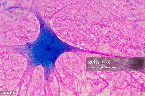
motor neuron (multipolar) with cell body and many processes (mostly dendrites), spinal cord (magnification x100). this multipolar motor neuron comes from the anterior (ventral) horn of the spinal cord grey matter. the stain is methylene blue and phloxine. - microphotographie immunofluorescente photos et images de collection
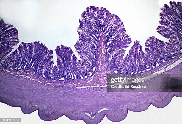
light micrograph of large intestine of human. mucosa, submucosa, muscularis, serosa, 10x at 35mm. goblet cells are visible in the mucosa. - microphotographie immunofluorescente photos et images de collection
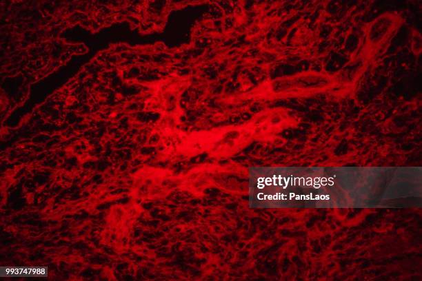
immunoflourescence d'une cellule musculaire lisse - microphotographie immunofluorescente photos et images de collection
inverted microscope viewing stem cells in flask with display of a fluorescent labeled cells - microphotographie immunofluorescente photos et images de collection
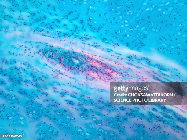
human hyaline cartilage, light micrograph - microphotographie immunofluorescente photos et images de collection
microscope of adenoid cystic carcinoma, rare type of cancer exist in many different body sites. - microphotographie immunofluorescente photos et images de collection
microscope of adenoid cystic carcinoma, rare type of cancer exist in many different body sites. - microphotographie immunofluorescente photos et images de collection
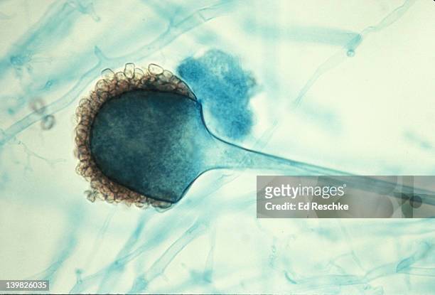
rhizopus (bread mold). sporanquim and spores. asexual reproduction. 100x - microphotographie immunofluorescente photos et images de collection
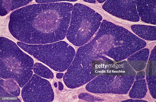
thymus. 10x shows: numerous lobules, cortex (darker with numerous lymphocytes), medulla (lighter). produces a hormone thymosin. - microphotographie immunofluorescente photos et images de collection
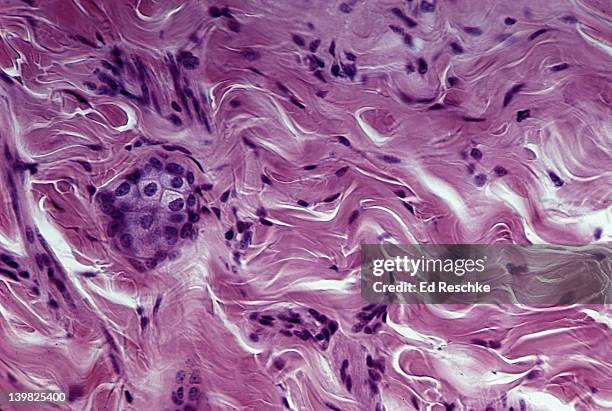
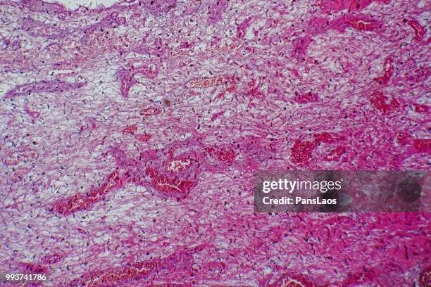
dense fibrous connective tissue (irregular); shows irregular collagenous fibers (pink) and fibroblast nuclei (purple); from the dermis of the skin - microphotographie immunofluorescente photos et images de collection
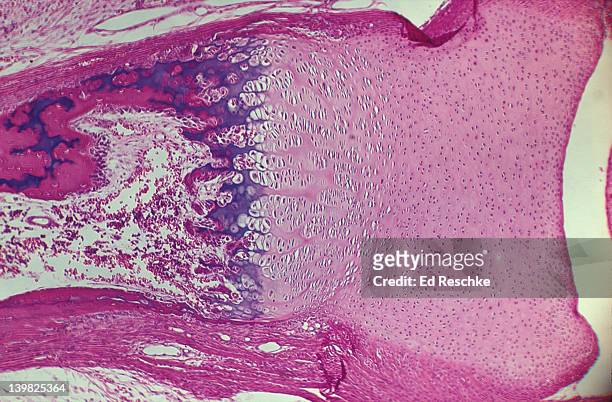
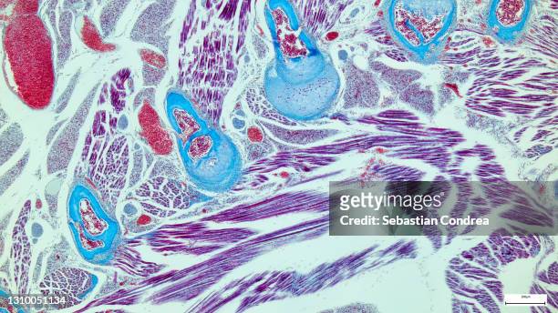
photomicrograph of endochondral ossification of developing bone in phalanx showing epiphyseal disk, hyaline cartilage, calcified cartilage and osteocytes. - microphotographie immunofluorescente photos et images de collection
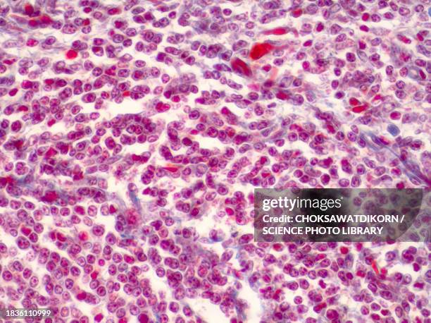
leukemia; myeloid, 250x at 35mm. this type of leukemia has its origin in the bone marrow (myeloid tissue). it involves a malignant proliferation of immature white blood cells. this action can crowd out production of rbc s and platelets leading to an - microphotographie immunofluorescente photos et images de collection
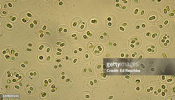
gloeocapsa, a cyanobacteria, photosynthetic, gram-negative, 100x at 35mm. formerly known as blue-green algae. prokaryotic cell. cells enclosed in layers of gelatinous material. - microphotographie immunofluorescente photos et images de collection
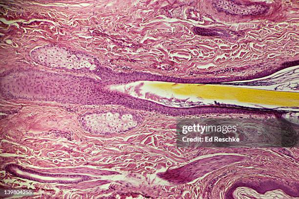
sebaceous glands and hair follicle of human scalp (magnification x25) h & e stain. showing hair follicle, hair, epidermis and dermis and arrector pili muscle - microphotographie immunofluorescente photos et images de collection
dicot stem. helianthus (sunflower) vascular bundles (ring), phloem, xylem, pith cortex, epidermis. 10x at 35mm. vascular bundles arranged in a ring. organization of primary tissues. - microphotographie immunofluorescente photos et images de collection
human scalp, shows: hair follicle, hair shaft, sebaceous glands, dermis & arrector pili muscle. 25x at 35mm - microphotographie immunofluorescente photos et images de collection
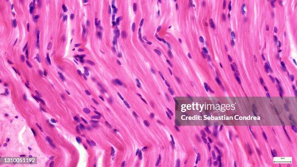
tendons have high tensile strength, light micrograph.immunofluorescent photomicrograph, organs samples, histological examination, histopathology on the microscope - microphotographie immunofluorescente photos et images de collection
sperm cells or spermatozoa, immunofluorescent photomicrograph, organs samples, histological examination, histopathology on the microscope - microphotographie immunofluorescente photos et images de collection
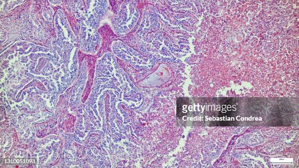
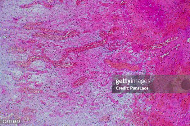
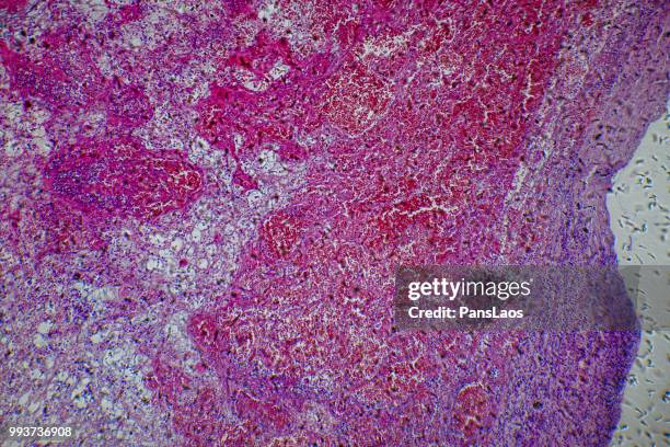
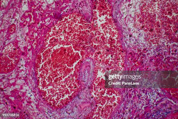
human lung biopsy under microscopy. immunofluorescent photomicrograph, organs samples, histological examination, histopathology on the microscope - microphotographie immunofluorescente photos et images de collection
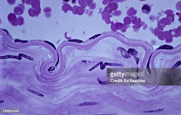
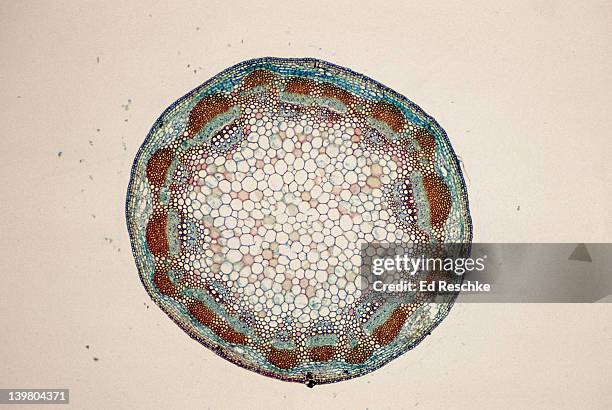
high resolution cross section of ascaris, giant roundworm, parasitic worm in humans. - microphotographie immunofluorescente photos et images de collection
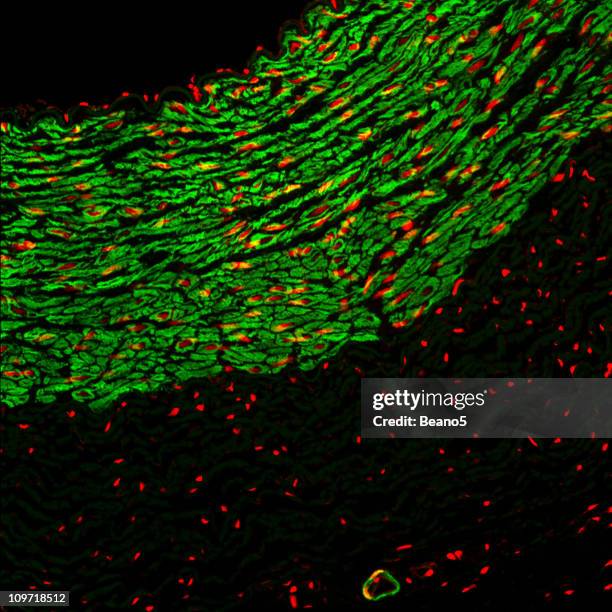
micrograph of rat brain. science cross section. immunofluorescent photomicrograph, organs samples, histological examination, histopathology on the microscope - microphotographie immunofluorescente photos et images de collection
immunofluorescent photomicrograph, organs samples, histological examination, histopathology on the microscope - microphotographie immunofluorescente photos et images de collection
human liver tissue under microscope view for education histology, human tissue - microphotographie immunofluorescente photos et images de collection